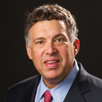
Interview with Roy S. Herbst, MD, PhD, and David L. Rimm, MD, PhD
At ASCO 2018, Oncology Practice Management (OPM) spoke with Roy S. Herbst, MD, PhD, Chief of Medical Oncology and Associate Director for Translational Research at Yale Cancer Center/Smilow Cancer Hospital, New Haven, CT, and David L. Rimm, MD, PhD, Professor of Pathology, Yale University School of Medicine, about the increasing role of biomarkers in the management of patients with lung cancer.
OPM: Molecular biomarkers have had a significant impact on cancer treatment, especially in immunotherapy, and especially in lung cancer. How has this affected your practices as an oncologist and a pathologist?
Dr Herbst: As an oncologist, I now don’t see patients until they’ve had biomarker testing. In lung cancer, it’s critical that we know the patient’s status for EGFR, ALK, ROS1, and RET mutations, as well as the PD-L1 status.
David and I work together at the Yale Cancer Center. Early on, David developed the PD-L1 assay, which is very important, because lung cancer treatment is now more personalized than ever. We need to know the PD-L1 status, and whether a patient has a molecular driver to be able to treat that patient properly. Even with the current chemotherapy combinations, it’s important to know the PD-L1 status.
Dr Rimm: This is an exciting time for pathology. We need to be ready to provide genetic information on the increasing numbers of new gene mutations. A few years ago, immunotherapy was a true breakthrough, and now we have immunohistochemistry (IHC) companion diagnostics for those new therapies. Roy and I have developed the FDA-approved companion diagnostic for PD-L1 status in lung cancer. We were one of the first centers to use the test routinely, and now most sites are using this test.
OPM: As a pathologist, is it becoming more difficult to incorporate the increasing array of biomarkers into your samples?
Dr Rimm: The specimens are getting smaller, and the demand for information from those small specimens is increasing. We can do that by having an efficient use of tissue, but we are often “stuck” with not enough tissue.
OPM: Where does the discussion between the oncologist and the pathologist occur? Is this at a tumor board? Is there a special committee? Something else?
Dr Herbst: Communication between the medical oncologist, the pathologist, as well as the surgeon, the pulmonologist, the radiologist, and the team obtaining the biopsy is critical. In lung cancer, we now treat all tumors in a personalized way, so it’s very important to know which markers are involved, and to have that information up front. This is an ever-changing field. Our techniques must evolve as new discoveries are made.
At Yale Cancer Center, we are fortunate to have many things that facilitate routine communication among the team, including tumor boards, where we present cases, which also includes a pathology review, radiology review, and then a clinical discussion. What makes a hospital such as ours very special is the research infrastructure.
I focus on lung cancer, because it’s what I know best, but we’re doing this in all areas. We have a lung cancer research working group, in which we meet every week to discuss what’s new in lung cancer, and what are the biomarkers. My goal is to be able to provide any patient the most up-to-date care, which includes testing for EGFR, ALK, ROS1, RET, MET, BRAF, and HER2 biomarkers. We have drugs for all these markers, but many are still in the investigational stage, which is also important to know.
We also have a second level of testing. We can test 136 genes using a next-generation sequencing (NGS) platform. We can also do whole-exome testing, if necessary. If the tumor mutational burden (TMB) is relevant, we can ask for NGS.
David works closely with us in the clinical and the research settings as part of our lung cancer research. We will move the field forward by developing those predictive markers for immunotherapy. David and I got to know each other when we were doing phase 1 clinical trials. We said we had to develop PD-L1 assays, and David developed several of those. He’s one of the best experts on how that works.
This also helped the clinical care: we can get a PD-L1 test for a patient with lung cancer quicker than we can do any other tests. This is important, because when drugs are approved based on a biomarker, or the biomarker helped to inform whether we should use combination drugs, we already have the information. It’s a wonderful collaboration, and there should be more pathologists who work like this. You need to think about how you’re collecting the tissue, how you will analyze the tissue, and multiplex the assays. Only a limited amount of tissue is being collected, so we need to prioritize how we use the tissue and multiplex it.
Dr Rimm: Roy and Lieping Chen run a specialized program for research excellence at Yale—our SPORE program, which is designed to be translational research. It allows us to take what is discovered in the lab and move it directly to the clinic for patient care.
This goes both ways—from patient care we work toward developing assays used in clinical care. And from the research side, we’re developing assays that can be tested in the clinical setting.
OPM: You had mentioned that biomarker testing is done at a different level. What biomarker tests do you routinely get for a patient with lung cancer? What would drive getting other tests, such as TMB or microsatellite instability (MSI), or a whole-exome analysis?
Dr Rimm: The standard has recently been published in the guidelines from the International Association for the Study of Lung Cancer. We want to make sure we’re doing everything according to the standards, which is testing for EGFR mutations.
We look at the sequence for the whole cytoplasmic domain to catch every possible mutation. Then we also test for ALK and ROS1 markers. Our routine 50-gene panel tests for more than that, including MET, RB-2, RET, and BRAF mutations. We test for several genes for every patient when the patient just comes in. So we’re meeting the guideline and exceeding it by testing all these other genes. Then we test for PD-L1. The guideline also says to test for PD-L1 in every patient. That’s the starting point, which is changing. In fact, even how we report the starting point is going to change after the ASCO 2018 meeting.
We heard in the plenary session about changing the cut point, so Roy and I will have to change our report. The panel we do for every patient is fluid, and it can respond to new developments in immunotherapy.
OPM: Where do these other markers—TMB or MSI—come into the picture?
Dr Rimm: MSI is interesting. That is, in a tumor with MSI or mismatch repair-deficient (dMMR), we could get a drug independent of whether it was prostate cancer, lung cancer, or another tumor. Now we have requests for MSI tests, but that is only for stage IV or high-grade cancers.
We don’t routinely test for MSI, except for colon cancer. But now we’re getting requests for MSI or dMMR testing for different cancer types. Pathologists are getting together to decide how we’re going to manage the requests for all tumor types instead of just one type. Any patients with an MSI or dMMR biomarker should be given the approved drug, and so we need to broaden our testing accordingly.
The TMB marker is a different story; it is still experimental. There’s been much hype around TMB, and some people in the oncology arena think TMB is “ready to go,” but it’s not. It’s been shown in several studies to be very promising. Perhaps the most exciting thing about TMB is that it’s complementary to the PD-L1 test. Once TMB is standardized and we can test for it, it will be the same at all cancer hospitals.
Those tests have to be rigorous and standardized at different sites. When standardized studies come out, as was the case 2 years ago for PD-L1, the PD-L1 test had broad adoption after the standardization. The same thing will likely happen with TMB. Once it’s standardized, we’ll see adoption, depending on the value of the test. Right now, the value of the test has only been shown in a couple of trials. It’s not very sensitive and specific; it’s about the same as PD-L1, but it’s different. Maybe we’ll do both tests to maximize the number of patients who can use the relevant drugs.
Dr Herbst: TMB is becoming more interesting by the day, and patients whose tumors don’t express PD-L1 can still have high TMB. The progression-free survival and the response rates to combinations of drugs, such as anti–PD-1 and CTLA-4 inhibitors, look promising.
Dr Rimm: I don’t want to bring up a TMB test unless I know that the TMB test that I do at Yale is the same test they do at other sites. We’re still not ready to do that.
Dr Herbst: There’s a national effort now to standardize TMB. It is tricky, because we don’t yet know how many genes to test for TMB, and how it correlates with treatments. It’s going to happen faster with immunotherapy, because all the tools are there. But it’s complicated, because we have to consider the tumor, as well as the tumor microenvironment.
I would predict that we’re going to personalize immunotherapy. As good as the data are in lung cancer, we’re still seeing most patients with lung cancer die of that disease. Only patients who have markers we have identified live 5 or 10 years with metastatic disease. We need to continue to search beyond TMB and PD-L1, to include the tumor microenvironment.
Dr Rimm: I agree that we have to keep our eye on phase 1 clinical trials and the drug pipeline. There will likely be other drugs that are very different from anti–PD-L1 that we see over the next few years. When we have more than 1 drug, we need tests that correspond to those drugs. We may soon have a tumor-infiltrating lymphocytes test that works across different drugs. We have assays ready in melanoma, and we’re going to start doing quantitative assessment of some lymphocytes. We’ll then move these to lung cancer or to other tumors, as needed.
The test is about the status of the patient, not about the status of the tumor. This tells us whether the patient is likely to respond to the treatment, or whether the patient should be triaged to another treatment. As more drugs become available, we’ll have more of these diagnostic tests.
OPM: We’ve heard that getting sufficient tissue for PD-L1 testing is a problem. Can you elaborate on this?
Dr Herbst: Sufficiency of tissue is a big issue. At a clinical trial recently, the group wanted 40 sections from a core biopsy. But if we provide all that, we wouldn’t have the tissue we need to do the test that is needed for immediate patient care. It is a problem that is going to require multiplexing and prioritization.
Dr Rimm: The amount of tissue is critical. In fact, because biopsies are small, we can make nucleic acids from them. If we make nucleic acids, we can’t test for PD-L1 or for other markers that indicate whether it is an adenocarcinoma or squamous-cell lung cancer. We have to triage the amount of tissue we get. If a clinical trial requires 40 sections, that exhausts the tissue.
As a pathology lab, we are also required to keep some diagnostic material. The patient may come back 2 years later, and we have to keep it so that we have enough tissue to review, and enough to do an NGS test.
OPM: What best practices can you share that nonacademic cancer centers or private practices can adopt to optimize biomarker testing, reporting, and application to the patient?
Dr Rimm: One of our best practices that is critical is trying to have rapid turnaround of the key information for the oncologist. When oncologists see that patient, we want them to already know the mutation status and what the PD-L1 status is.
Those are the 2 big things in lung cancer—the mutation status and the PD-L1 status. Figuring out a way to get those turned around as quickly as possible has been our goal. In terms of timing, we want the PD-L1 data to be turned around in 72 hours. We’d like the mutation status to be turned around in 10 days. Sometimes it takes us 14 days, but from the pathology perspective, we have to give that information to the clinician so he or she can best manage the patient.
Dr Herbst: I would agree. Best practices are very important. We’re very fortunate at Yale to have much of this in-house. Most community centers will be outsourcing this work, which is fine as long as they use a reliable place that provides the results in the set period of time. But more important is the interpretation—knowing what to do with the results. Why send a test if you’re not going to act on the results?
At tumor board meetings, when asked to understand what the markers mean, or how to use them, I would expect that the group that does your marker analysis also has a way of communicating to you what it means.
Let’s say you do a TMB analysis. You may want to call someone at the center and ask how to interpret this. If they say, “Well, we don’t have a lot of tissue, but we’re going to micro-dissect it, and we’ll make it work,” and explain some of the physical characteristics, that can help. The more communication you have, the better. Education and communication are key.
Always remember that there’s a patient on the other end waiting for the result that will affect what treatment the patient gets. Tissue is very precious. We’ve got to use it carefully. If we send it out for a test, we have to ensure we have enough left to do the test we need to treat that patient with the best available therapy, in real time.
OPM: Do you start treatment for any patient before you get the pathology results back?
Dr Herbst: I almost always wait for the pathology results to come back before I’d start treatment; it’s important to know whether it’s a squamous-cell or a nonsquamous tumor.
As far as molecular studies, if it’s a patient with a nonsquamous-cell tumor, I want to know what the driver mutations are before I proceed. The same is true for patients with squamous-cell tumor. Not everyone does that, because sometimes the histology is not clear.
That said, in rare situations you could start with chemotherapy, or now perhaps with chemotherapy plus immunotherapy, but I don’t like that. I would try to wait to get the pathology results. There are very few true emergencies when chemotherapy is going to cure that emergency, in my opinion.
I’ve had cases where the staff were all anxious to get started, when the PET scan looked awful. If the patient has a performance status 0 or 1, I’ll wait. Occasionally, you have to start sooner. The way to avoid this is to get the testing so routine that it gets out as quickly as possible so that you get back at least the driver mutation status and the PD-L1 status before beginning therapy.
OPM: Are liquid biopsies of any value?
Dr Rimm: Liquid biopsies are interesting: they allow us to get more tissue, because we just draw more blood, and we can get it at multiple time points. The problem with liquid biopsies is that there are hardly any data involved. It’s like looking at smoke instead of fire. Sometimes the smoke tells you what’s going on, but not always.
Studies are now evaluating the value of liquid biopsy. In some targeted therapies, there’s already clear use for liquid biopsies. Even the 2018 recommendations from our pathology and oncology societies already recommend to use liquid biopsy when we can’t acquire tissue.
Dr Herbst: I use a liquid biopsy in the clinic when I want something quickly. It can be done in real time. If it’s negative, though, I still need to use the tissue biopsy. Targeted liquid biopsies can come back quickly. When someone has a disease such as recurrent EGFR-mutated lung cancer, there are no easy sites to biopsy tissue. What if it’s in the bone or something like that?
Liquid biopsies can be very useful. I think they are going to be the future, and the technology keeps getting better every year.
OPM: If the liquid biopsy is positive for a predictive biomarker, can you act on that?
Dr Herbst: If it’s positive, yes. If it’s negative, then we have to keep looking.
Dr Rimm: Liquid biopsies can be better if we know what gene we’re looking for. In the case of disease recurrence, we’re looking for just a handful of genes.
Even looking for TMB from liquid biopsies is more challenging than looking for EGFR mutations. There are only 7 or 12 mutations we look for with liquid biopsy. Our company is developing tests to allow us to get the results in 24 hours. That’s a real service to our oncologist colleagues.
OPM: Thank you both for this exceedingly informative conversation.